Showing posts with label Neuroscience. Show all posts

Sunday, April 21, 2013

Stem Cell Transplant Restores Memory, Learning in Mice

 For the first time, human embryonic stem cells have been transformed into nerve cells that helped mice regain the ability to learn and remember.
A study at the University of Wisconsin-Madison is the first to show that human stem cells can successfully implant themselves in the brain and then heal neurological deficits, says senior author Su-Chun Zhang, a professor of neuroscience and neurology.  Once inside the mouse brain, the implanted stem cells formed two common, vital types of neurons, which communicate with the chemicals GABA or acetylcholine. "These two neuron types are involved in many kinds of human behavior, emotions, learning, memory, addiction and many other psychiatric issues," says Zhang.

Yan Liu, et al.
Medial ganglionic eminence–like cells derived from human embryonic stem cells correct learning and memory deficits. Nature Biotechnology, 2013; DOI:10.1038/nbt.2565

Monday, April 15, 2013

Brain Development Is Guided by Junk DNA That Isn't Really Junk

 Specific DNA once dismissed as junk plays an important role in brain development and might be involved in several devastating neurological diseases, UC San Francisco scientists have found.
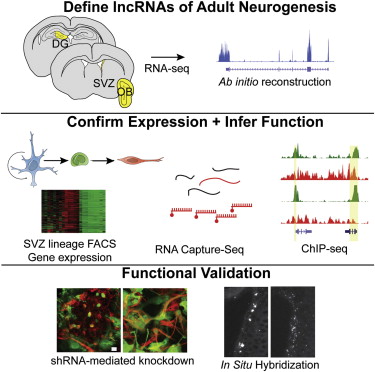
Their discovery in mice is likely to further fuel a recent scramble by researchers to identify roles for long-neglected bits of DNA within the genomes of mice and humans alike. While researchers have been busy exploring the roles of proteins encoded by the genes identified in various genome projects, most DNA is not in genes. This so-called junk DNA has largely been pushed aside and neglected in the wake of genomic gene discoveries, the UCSF scientists said. In their own research, the UCSF team studies molecules called long noncoding RNA (lncRNA, often pronounced as "link" RNA), which are made from DNA templates in the same way as RNA from genes.
Science Daily, Apr. 15, 2013

Alexander D. Ramos, et al.
Integration of Genome-wide Approaches Identifies lncRNAs of Adult Neural Stem Cells and Their Progeny In Vivo.
Cell Stem Cell, 2013 DOI:10.1016/j.stem.2013.03.003

Friday, April 12, 2013

Deep Homology of Arthropod Central Complex and Vertebrate Basal Ganglia

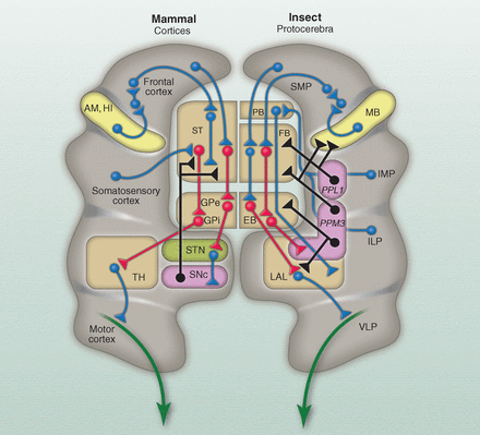
Similarities of brain structure, function, and behavior are usually ascribed to convergent evolution. In their review, Strausfeld and Hirth (p. 157) identify multiple commonalities shared by vertebrate basal ganglia and a system of forebrain centers in arthropods called the central complex. The authors conclude that circuits essential to behavioral choice originated very early across phyla.

Nicholas J. Strausfeld, et al
Science 12 April 2013:
Vol. 340 no. 6129 pp. 157-161 , DOI: 10.1126/science.1231828

Thursday, April 11, 2013

See-through brains clarify connections

A chemical treatment that turns whole organs transparent offers a big boost to the field of ‘connectomics’ — the push to map the brain’s fiendishly complicated wiring. Scientists could use the technique to view large networks of neurons with unprecedented ease and accuracy. The technology also opens up new research avenues for old brains that were saved from patients and healthy donors.

Helen Shen
Nature 496, 151 (11 April 2013) doi:10.1038/496151a

Kwanghun Chung, et al
Structural and molecular interrogation of intact biological systems
Nature (2013) doi:10.1038/nature12107

Thursday, April 4, 2013

A 'Light Switch' in Brain Illuminates Neural Networks

 There are cells in your brain that recognize very specific places, and have that as one of their main jobs. These cells, called place cells, are found in an area behind your temple called the hippocampus. While these cells must be sent information from nearby cells to do their job, so far no one has been able to determine exactly what kind of nerve cells, or neurons, work with place cells to craft the code they create for each location. Neurons come in many different types with specialized functions. Some respond to edges and borders, others to specific locations, others act like a compass and react to which way you turn your head.
Science News Apr. 4, 2013 

Sheng-Jia Zhang*,†, Jing Ye*, Chenglin Miao, Albert Tsao, Ignas Cerniauskas, Debora Ledergerber, May-Britt Moser, Edvard I. Moser. Optogenetic Dissection of Entorhinal-Hippocampal Functional ConnectivityScience, 5 April 2013: Vol. 340 no. 6128 DOI: 10.1126/science.1232627

Thursday, March 28, 2013

Systematic Analysis of Neural Projections Reveals Clonal Composition of the Drosophila Brain

During development neurons are generated by sequential divisions of neural stem cells, or neuroblasts. In the insect brain progeny of certain stem cells form lineage-specific sets of projections that arborize in distinct brain regions, called clonal units. Though this raises the possibility that the entire neural network in the brain might be organized in a clone-dependent fashion, only a small portion of clones has been identified.

Our study showed that the insect brain is formed by a composition of cell-lineage-dependent modules. Clonal analysis reveals organized architecture even in those neuropils without obvious structural landmarks.

Current Biology, online 28 March 2013
http://dx.doi.org/10.1016/j.cub.2013.03.015


Thursday, March 21, 2013

Forgetting in C. elegans Is Accelerated by Neuronal Communication via the TIR-1/JNK-1 Pathway

The control of memory retention is important for proper responses to constantly changing environments, but the regulatory mechanisms underlying forgetting have not been fully elucidated. Our genetic analyses in C. elegans revealed that mutants of the TIR-1/JNK-1 pathway exhibited prolonged retention of olfactory adaptation and salt chemotaxis learning. In olfactory adaptation, conditioning induces attenuation of odor-evoked Ca2+ responses in olfactory neurons, and this attenuation is prolonged in the TIR-1/JNK-1-pathway mutant animals. We also found that a pair of neurons in which the pathway functions is required for the acceleration of forgetting, but not for sensation or adaptation, in wild-type animals. In addition, the neurosecretion from these cells is important for the acceleration of forgetting. Therefore, we propose that these neurons accelerate forgetting through the TIR-1/JNK-1 pathway by sending signals that directly or indirectly stimulate forgetting.

Akitoshi Inoue, et al
Cell Reports, Volume 3, Issue 3, 808-819, 21 March 2013


Friday, March 15, 2013

Epigenetic mechanisms in the development and maintenance of dopaminergic neurons

Mesodiencephalic dopaminergic (mdDA) neurons are located in the ventral mesodiencephalon and are involved in psychiatric disorders and severely affected in neurodegenerative diseases such as Parkinson’s disease. mdDA neuronal development has received much attention in the last 15 years and many transcription factors involved in mdDA specification have been discovered. More recently however, the impact of epigenetic regulation has come into focus, and it’s emerging that the processes of histone modification and DNA methylation form the basis of genetic switches that operate during mdDA development. Here, we review the epigenetic control of mdDA development, maturation and maintenance. As we highlight, epigenetic mechanisms play a pivotal role in all of these processes and the knowledge gathered from studying epigenetics in these contexts may aid our understanding of mdDA-related pathologies.

Hendrikus J. van Heesbeen, et al
Development140, 1159-1169.   March 15, 2013
doi:10.1242/dev.089359  

Saturday, January 26, 2013

Developmental processes in face perception

Understanding the developmental origins of face recognition has been the goal of many studies of various approaches. Contributions of experience-expectant mechanisms (early component), like perceptual narrowing, and lifetime experience (late component) to face processing remain elusive. By investigating captive chimpanzees of varying age, a rare case of a species with lifelong exposure to non-conspecific faces at distinctive levels of experience, we can disentangle developmental components in face recognition. We found an advantage in discriminating chimpanzee above human faces in young chimpanzees, reflecting a predominant contribution of an early component that drives the perceptual system towards the conspecific morphology, and an advantage for human above chimpanzee faces in old chimpanzees, reflecting a predominant late component that shapes the perceptual system along the critical dimensions of the face exposed to. We simulate the contribution of early and late components using computational modeling and mathematically describe the underlying functions.

Christoph D. Dahl, et al.
Published 09 January 2013

Wednesday, January 23, 2013

Action Plan: Making Brain-Controlled Prosthetics That Can Open a Clothespin

Brain-controlled interfaces have advanced dramatically during the past decade. But more work needs to be done before this technology begins to approximate the natural movements of a fully functioning arm or hand. An attempt to replicate the full range of movement—and the cognitive chain of events from thought to action—has now begun as a research collaboration among the California Institute of Technology, Johns Hopkins University Applied Physics Laboratory, the University of Southern California and Rancho Los Amigos National Rehabilitation Center. These institutions are seeking a few recruits to be fitted with a $500,000 robotic limb.

By Gary Stix,  January 23, 2013, Scientific American


Billion-euro brain simulation and graphene projects win European funds

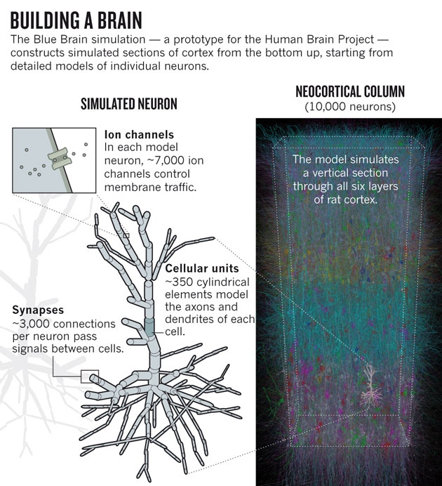
The European Commission has selected the two research proposals it will fund to the tune of half-a-billion euros each after a two-year, high-profile contest. The Human Brain Project, led by neuroscientist Henry Markram at the Swiss Federal Institute of Technology (EPFL) in Lausanne, plans to simulate everything known about the human brain in a supercomputer — a breathtaking ambition that has been met with some scepticism (See “Brain in a box”).

NATURE | BREAKING NEWS, 23 January 2013
Nature 482, 456–458 (23 February 2012) doi:10.1038/482456a


Sunday, January 20, 2013

Grid cells require excitatory drive from the hippocampus

To determine how hippocampal backprojections influence spatially periodic firing in grid cells, the author recorded neural activity in the medial entorhinal cortex (MEC) of rats after temporary inactivation of the hippocampus. They report two major changes in entorhinal grid cells. First, hippocampal inactivation gradually and selectively extinguished the grid pattern. Second, the same grid cells that lost their grid fields acquired substantial tuning to the direction of the rat's head. This transition in firing properties was contingent on a drop in the average firing rate of the grid cells and could be replicated by the removal of an external excitatory drive in an attractor network model in which grid structure emerges by velocity-dependent translation of activity across a network with inhibitory connections. These results point to excitatory drive from the hippocampus, and possibly other regions, as one prerequisite for the formation and translocation of grid patterns in the MEC.

Tora Bonnevie, et al.
Nature Neuroscience (2013) doi:10.1038/nn.3311, Published online 20 January 2013


One Form of Neuron Turned Into Another in Brain

 A new finding by Harvard stem cell biologists turns one of the basics of neurobiology on its head -- demonstrating that it is possible to turn one type of already differentiated neuron into another within the brain. The discovery by Paola Arlotta and Caroline Rouaux "tells you that maybe the brain is not as immutable as we always thought, because at least during an early window of time one can reprogram the identity of one neuronal class into another," said Arlotta.

Caroline Rouaux, Paola Arlotta.
Direct lineage reprogramming of post-mitotic callosal neurons into corticofugal neurons in vivo. 

Recurrent inhibitory circuitry as a mechanism for grid formation


Grid cells in layer II of the medial entorhinal cortex form a principal component of the mammalian neural representation of space. The firing pattern of a single grid cell has been hypothesized to be generated through attractor dynamics in a network with a specific local connectivity including both excitatory and inhibitory connections. However, experimental evidence supporting the presence of such connectivity among grid cells in layer II is limited. Here we report recordings from more than 600 neuron pairs in rat entorhinal slices, demonstrating that stellate cells, the principal cell type in the layer II grid network, are mainly interconnected via inhibitory interneurons. Using a model attractor network, we demonstrate that stable grid firing can emerge from a simple recurrent inhibitory network. Our findings thus suggest that the observed inhibitory microcircuitry between stellate cells is sufficient to generate grid-cell firing patterns in layer II of the medial entorhinal cortex.

Jonathan J Couey, et al.
Nature Neuroscience (2013) doi:10.1038/nn.3310, Published online 20 January 2013


Thursday, January 17, 2013

Human brain evolution: transcripts, metabolites and their regulators

What evolutionary events led to the emergence of human cognition? Although the genetic differences separating modern humans from both non-human primates (for example, chimpanzees) and archaic hominins (Neanderthals and Denisovans) are known, linking human-specific mutations to the cognitive phenotype remains a challenge.  The new strategy is to focus on human-specific changes at the level of intermediate phenotypes, such as gene expression and metabolism, in conjunction with evolutionary changes in gene regulation involving transcription factors, microRNA and proximal regulatory elements. In this Review the authors show how this strategy has yielded some of the first hints about the mechanisms of human cognition.


Discrete genetic modules are responsible for complex burrow evolution in Peromyscus mice

The genetics of behavioural differences between closely related species are less well understood than the genetics of morphological differences. Many animals build elaborate structures — such as hives, nests and burrows — that 'evolve' as natural selection acts on the behaviour of their builders. This study uses an example of this phenomenon to tackle the question of whether complex behaviours evolve through one or few genetic changes that each influence many aspects of behaviour, or by accumulation of several genetic changes that generate behavioural complexity only when combined. Hopi Hoekstra and colleagues show that the complex burrows created by oldfield mice are governed by several genetic modules, each controlling an aspect of burrow size or shape. This modularity in burrow architecture suggests that complex behaviour may result from the combination of genetically determined behaviours that have accumulated over time.

Jesse N. Weber, et al.

Why Wolves Are Forever Wild, but Dogs Can Be Tamed

Dogs and wolves are genetically so similar, it's been difficult for biologists to understand why wolves remain fiercely wild, while dogs can gladly become "man's best friend." Now, doctoral research by evolutionary biologist Kathryn Lord at the University of Massachusetts Amherst suggests the different behaviors are related to the animals' earliest sensory experiences and the critical period of socialization. Details appear in the current issue of Ethology.

When the socialization window is open, wolf and dog pups begin walking and exploring without fear and will retain familiarity throughout their lives with those things they contact. Domestic dogs can be introduced to humans, horses and even cats at this stage and be comfortable with them forever. But as the period progresses, fear increases and after the window closes, new sights, sounds and smells will elicit a fear response.  Through observations, Lord confirmed that both wolf pups and dogs develop the sense of smell at age two weeks, hearing at four weeks and vision by age six weeks on average. However, these two subspecies enter the critical period of socialization at different ages. Dogs begin the period at four weeks, while wolves begin at two weeks. Therefore, how each subspecies experiences the world during that all-important month is extremely different, and likely leads to different developmental paths, she says.
Science Daily, Jan. 17, 2013

Kathryn Lord.
A Comparison of the Sensory Development of Wolves (Canis lupus lupus) and Dogs (Canis lupus familiaris). 
Ethology, 2013; 119 (2): 110 DOI:10.1111/eth.12044


Wednesday, January 16, 2013

Light Switch Inside Brain: Laser Controls Individual Nerve Cells in Mouse

Activating and deactivating individual nerve cells in the brain is something many neuroscientists wish they could do, as it would help them to better understand how the brain works.  Scientists in Freiburg and Basel, Switzerland, have developed an implant that is able to genetically modify specific nerve cells, control them with light stimuli, and measure their electrical activity all at the same time. This novel 3-in-1 tool paves the way for completely new experiments in neurobiology.

A polymer-based neural microimplant for optogenetic applications: design and first in vivo study
Birthe Rubehn, et al.
Lab Chip, 2013, Advance Article
DOI: 10.1039/C2LC40874K, First published on the web 03 Jan 2013


Monday, January 14, 2013

Memory on time

Considerable recent work has shown that the hippocampus is critical for remembering the order of events in distinct experiences, a defining feature of episodic memory. Correspondingly, hippocampal neuronal activity can ‘replay’ sequential events in memories and hippocampal neuronal ensembles represent a gradually changing temporal context signal. Most strikingly, single hippocampal neurons – called time cells – encode moments in temporally structured experiences much as the well-known place cells encode locations in spatially structured experiences. These observations bridge largely disconnected literatures on the role of the hippocampus in episodic memory and spatial mapping, and suggest that the fundamental function of the hippocampus is to establish spatio-temporal frameworks for organizing memories.

Howard Eichenbaum
Trends in Cognitive Sciences, 14 January 2013
http://www.sciencedirect.com/science/article/pii/S1364661312002896

New Implant Replaces Impaired Middle Ear

Functionally deaf patients can gain normal hearing with a new implant that replaces the middle ear. The unique invention from the Chalmers University of Technology has been approved for a clinical study. The first operation was performed on a patient in December 2012.